Listing ID #381491
Company Information
Ask for more detail from the seller
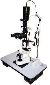
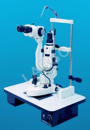
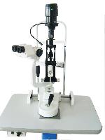
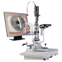
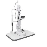
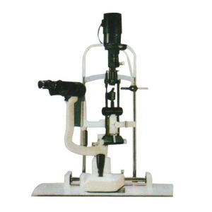
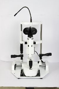
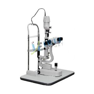
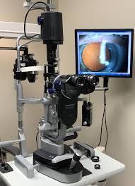
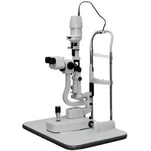
Contact Supplierwe are reckoned as one of the leading slit lamp microscope manufacturers, exporters and suppliers in haryana. The slit lamp is essentially a simple and under-used piece of equipment. The slit lamp consists of an illumination system and a binocular observation system, which when correctly aligned will result in a coincidental focus of the slit and microscope. illumination system a short focus projector projecting an image of the illuminated slit aperture on to the eye. This part of the system should be flexible to allow various sizes and shape of slit beam. Usually a rheostat is incorporated and the lamp house can be rotated. Neutral density, cobalt blue and red free filters are usually available, and occasionally a diffuser and polarizer using the slit lamp commence the examination using the 10x eyepieces and the lower powered objective i.e. 1x. Use the lowest voltage setting on the transformer. Select the longest slit length by means of the appropriate lever (17). Adjust the chin rest by means of control 27 so that the patient's eyes are approximately level with the black marker on the side of the head rest. Adjust the height of the slit lamp until the slit beam is centered vertically on the patient's eye. Focus the slit beam on the eye by moving the joystick (1) either towards or away from the patient. Coarse positioning can be effected without using the microscope but critical focusing should be carried out whilst viewing through the microscope. either the slit width is varied by rotating the left hand or right hand knurled control 10. To vary the angle between illumination and microscope use one or other of these same controls as handles. The slit should be set primarily in the vertical position, but any desired inclination can be achieved by means of the ball handle 15 (notches at 45°, 90°, 135°; stops at 0o and 180o). By tripping the latch 11 and tilting the slit lamp column, the beam can be introduced from as much as 20° below the horizontal. This is mainly used for carrying out gonioscopy. for observation by sclerotic scatter or other dissociated forms of examination, the centering screw 13 is loosened, so that the slit image can be moved away from the center of the field of observation. The image is centered again by tightening the screw.


Connect with us