


Individual (Sole proprietorship)
2022
6 - 20
Below Rs. 0.5 Crore Approx.
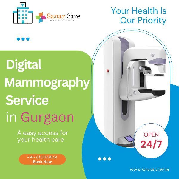
Mammography is a specialized medical imaging technique that uses low-dose X-rays to examine breast tissue for early detection and diagnosis of breast diseases, particularly breast cancer. It is a non-invasive procedure designed to identify abnormal changes, such as lumps, calcifications, or other irregularities, that may not be felt during a physical exam. Mammograms are commonly performed as part of routine breast cancer screening for women, especially those over the age of 40 or those at higher risk due to family history or genetic predisposition. The procedure involves compressing the breast between two plates to spread the tissue evenly, allowing for clearer imaging. Mammography plays a critical role in early detection, improving treatment outcomes and reducing breast cancer mortality rates. Advanced techniques, such as digital mammography and 3D tomosynthesis, provide enhanced image quality and more precise evaluation.
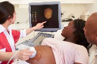
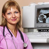
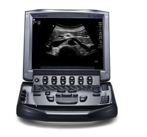
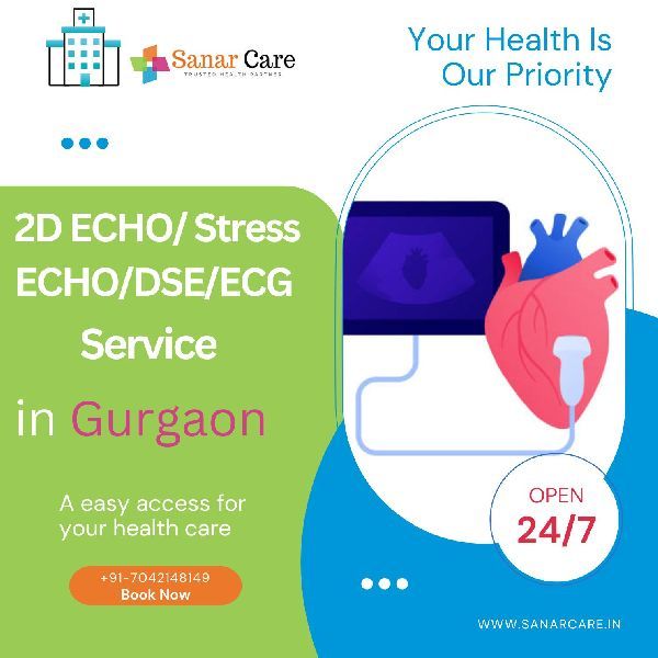
Ultrasound , also known as sonography , is a non-invasive imaging technique that uses high-frequency sound waves to create real-time images of internal organs, tissues, and blood flow. It is widely used for diagnostic purposes, guiding procedures, and monitoring medical conditions. During an ultrasound, a handheld device called a transducer is placed on the skin or inside the body, emitting sound waves that bounce off structures and create images displayed on a monitor. Common applications include examining the abdomen, pelvis, heart (echocardiography), blood vessels (Doppler ultrasound), and monitoring pregnancy to assess fetal development. Ultrasound is safe, painless, and does not use ionizing radiation, making it suitable for a broad range of patients, including pregnant women and children. Its portability and real-time imaging capabilities make it an essential tool in modern medicine.
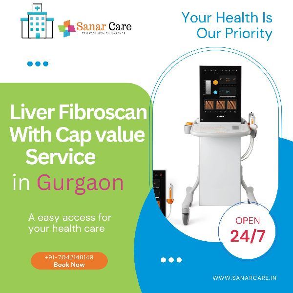
FibroScan is a non-invasive diagnostic tool used to assess liver health by measuring liver stiffness and fat content through transient elastography. It provides a quick and painless alternative to liver biopsy, taking only 5–10 minutes to deliver immediate results. By evaluating liver stiffness, FibroScan identifies the degree of fibrosis (scarring), while its controlled attenuation parameter (CAP) measures liver fat levels to detect conditions like fatty liver disease. Commonly used for managing chronic liver conditions such as hepatitis, alcoholic liver disease, and non-alcoholic fatty liver disease (NAFLD), it is safe, repeatable, and effective for monitoring liver health over time. However, factors like obesity or inflammation may influence its accuracy, and it is not designed to detect structural abnormalities such as tumors.
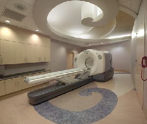
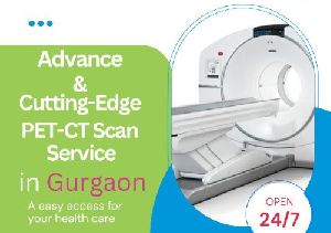
A PET CT scan (Positron Emission Tomography - Computed Tomography) is a powerful imaging technique that combines two advanced scans— PET and CT —into one procedure, providing highly detailed and functional information about the body. PET Scan : This part of the scan involves injecting a small amount of a radioactive tracer into the body. The tracer is absorbed by the cells, and the PET scanner detects radiation emitted by the tracer to create images showing the metabolic activity of tissues and organs. PET scans are particularly useful for detecting cancer, assessing how well cancer treatments are working, and identifying areas of abnormal brain or heart function. CT Scan : The CT component uses X-rays to take detailed cross-sectional images of the body’s internal structures, helping to localize and pinpoint the area of abnormality. By combining these two technologies, a PET CT scan provides detailed anatomical and functional images that allow doctors to assess not only the size and shape of a tumor or abnormality (from the CT part) but also how active it is metabolically (from the PET part). This makes PET CT scans extremely valuable in cancer detection, staging, and monitoring treatment effectiveness, as well as in cardiac and neurological evaluations. The procedure involves a small amount of radiation but is generally safe and offers precise diagnostic information.
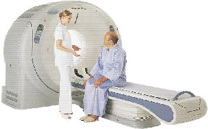
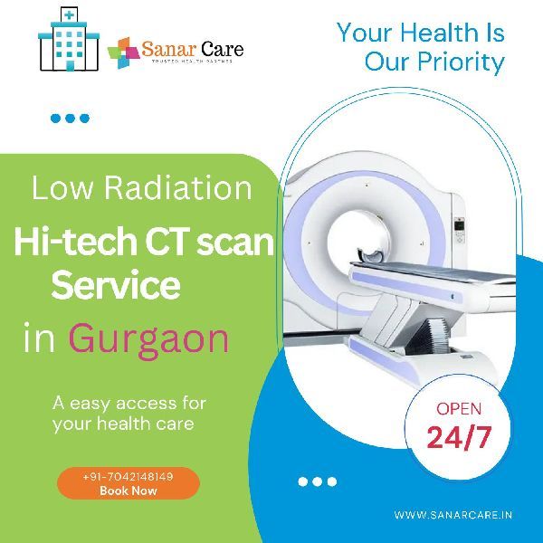
A CT scan (Computed Tomography scan) is a non-invasive imaging procedure that uses X-rays and advanced computer technology to create detailed cross-sectional images of the body. It provides highly accurate visualizations of internal organs, bones, blood vessels, and soft tissues, making it a vital diagnostic tool for detecting injuries, infections, tumors, and other abnormalities. During the scan, the patient lies on a motorized table that moves through a large, doughnut-shaped machine, which rotates to capture multiple X-ray images from different angles. These images are then processed by a computer to produce detailed 2D or 3D views. CT scans are commonly used in emergencies, preoperative planning, cancer diagnosis, and monitoring treatment progress. In some cases, a contrast dye may be administered orally or intravenously to enhance image clarity. While CT scans are quick and effective, they do involve exposure to a small amount of radiation, which is carefully managed to ensure patient safety.
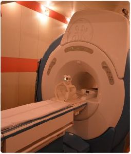
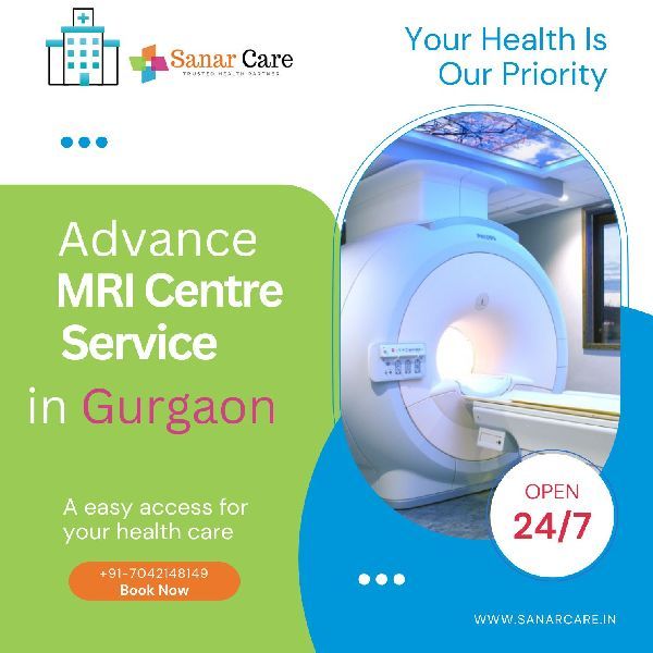
MRI (Magnetic Resonance Imaging) is an advanced, non-invasive diagnostic imaging technique that uses powerful magnets, radio waves, and a computer to create detailed images of the internal structures of the body. Unlike X-rays or CT scans, MRI does not use ionizing radiation, making it a safer option for long-term imaging needs. It is particularly effective for visualizing soft tissues, such as the brain, spinal cord, joints, muscles, and organs, as well as detecting abnormalities like tumors, inflammation, or injuries. During the procedure, the patient lies on a motorized table that slides into a cylindrical scanner. The strong magnetic field and radio waves interact with hydrogen atoms in the body to produce precise, high-resolution images. Specialized MRI techniques, such as contrast-enhanced MRI, functional MRI (fMRI), and cardiac MRI, allow for even more detailed evaluations. MRI is widely used in neurology, orthopedics, cardiology, and oncology for diagnosis and treatment planning. Though the process is painless, patients with claustrophobia or metal implants should inform their healthcare provider to ensure safety and comfort during the scan.
Share your thoughts with other customers for Sanar Care Diagnostic Centre
Add Review