High-precision delayed point-to-point dynamic receiving and focusing
Superwide frequency imaging
Adaptive image optimization
Adaptive vascular imaging
Adaptive Doppler imaging
THI
Applicable for the whole body, including but not limited to abdomen, Gyn., Obs., Cardiology, Vasuclar, Urology, Small organs, Mammary gland, paediatrics and neonatus, fetal cardiac imaging, puncture.
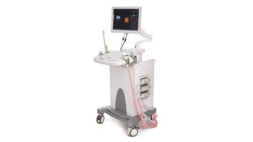
Integrative network ultrasonic workstation. Available for DICOM.
Scanning modes :B, M, CFM, PDI, PW, THI
Image processing technologies
Acoustic beam processing: full digital multi-beam former, real-time point-to-point dynamic receiving and focusing, continuous dynamic focusing, real-time dynamic variable aperture imaging, real-time dynamic acoustic beam apodization, dynamic filtering, dynamic frequency scanning.
Image pretreatment
Overall gain: 0~100 adjustable
TGC:8 TGC sliders
Gain control:B+M adjustable, CFM adjustable, PDI adjustable, PW adjustable
Acoustic output: Low, middle, high adjustable
Grey levels: grade 0 to 15 adjustable
Digital channels: 32
High-precision digital continuous beam former
Edge enhancement 5 grades adjustable
Frame average 6 grades adjustable
Line average 3 grades adjustable
Gamma correction grade 0 to 30 adjustable
Contrast: 0~100
Brightness: -50~+50
Scanning parameters
B mode
Acoustical power: Low, middle, high adjustable
B gains: 0-100
Danymic range: 120dB
M mode
M acoustical power: Low, middle, high adjustable
B gains: 0-100
M scanning speed: Low, middle, high adjustable
M sampling line visible & adjustable
CFM color Doppler mode
CFM gain: 0-100
CFM wall filtering: 4 steps
CFM baseline adjustment: 9 steps
CFM frame average: 4 steps
Maximum PRF:14kHz
Minimum PRF:0.5kHz
CDE Power Doppler mode
CDE gain: 0-100
PD frame average: 4 steps
PD wall filtering: 4 steps
Maximum PRF:14kHz
Minimum PRF:0.5kHz
PW Spectral Doppler mode
PW gain:0-100
PW dynamic range: 4 steps
PW wall filtering: 4 steps
PW color
Digital wall filtering: 4 steps
PW scanning speed: 4 steps (120,180,240,300)
Sampling volume width: 1-15mm
Angle correction
Baseline adjustment: 9 steps
Maximum PRF(speed):15kHz
Minimum PRF(speed):1kHz
Maximum detectable speed:21m/s(7.18m/s)
Minimum detectable speed:0.05cm/s
Image display
256 gray scale
Grey histogram display
Image rotation : left / right, up / down, 90oC
Depth : 3 to 24 cm (22 grades) depending on probes
Emit focus : maximum 8 focus points (depending on probe types and depth)
Dynamic range : ≥120dB (visible & adjustable)
Real-time zoom : 6 times zoon in, zoom rate adjustable
M mode speed : 3 grades adjustable
Angle change : 3 angles (available for convex probe)
Display TGC curve
Video output : auto-select NTSC or PAL video format
Screen display information
Header information area: hospital log, hospital name, system date and time, patient information, TI/MI value, probe model and current mode.
Menu adjustment area: image adjustment and optimization; pseudo colors, rotation and reverse; dynamic range, frame average, linear average, linear density, acoustic power of image real-time scanning; remark texts of current mode; body mark of current mode;basic measurement and applicable measurement items of current model under frozen condition.
Parameter and measurement area: parameters of current image scanning; related measurement and calculation results when measuring.
Image area: to display images under each mode and measurements and each texts, arrows.
Caliper area: to display depth reference caliper, grey grade caliper, CFM or power's speed caliper; TGC curve displays or hides automatically according to parameter setup.
Record area: to display current image and status messages being saved in video buffer in live condition; to display the saved total frames and status message of current frame under frozon condition.
Menu status area: to display the status of related parameters in both live and frozon conditions.
B mode normal measurements: distance, circumrence (Ellipse & method of loci), area (Ellipse & method of loci), volume (Ellipse, method of loci and two-planes models), ratio and angle.
M mode normal measurements: distance, time, slope and heart rate.
PW mode normal measurements: time, heart rate, velocity, acceleration, resistance index.
CFM、PDI mode normal measurements:distance, circumrence (Ellipse & method of loci), area (Ellipse & method of loci), volume (Ellipse, method of loci and two-planes models), ratio and angle.
Gynecological measurement: uterus (uterus, cervix and endometria), ovarian volume and left & right follicle.
Cardialogy measurement: applicable measurements for aortic, aortic vaivem, left atrium, LV, mitral vaive, right atrium, right ventricle, circulatory system.
Excellent measurement and calculation programs for General, Obstetrics, Gynecology, Urology, Small Parts, Cardiac, Skeletal & Muscles Vascular examinations especially on vessels, Blood
" Ultrasound Scanner Machines will be Sold & Purchased strictly as per the applicable Guidelines of PC & PNDT."